|
Zebrafish Embryo Datasets
|
|
Details
Description
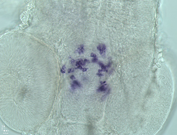
This dataset contains 35 zebrafish embryo stacks scanned under 20X magnification through wide field microscope. In the dataset, there are 25 stacks for training and 10 stacks for testing. The volume (in voxels) of each stack are 1024 ∗ 1344 ∗ z, and different stacks have different values of z. The spatial resolution is 3µm and the axial resolution is 1.5µm. The magnification of the objective is 20X, and the numerical aperture (NA) is 0.7. For each of the stack in this dataset, one professional observers label all centres of tyrosine hydroxylase (TH) cells (see reference [1]) using ’Point Picker’ [2], a plugin of the free JAVA application ’ImageJ’ 1.49j [3]. All the labelled points in one stack are saved in one .txt file. References [1] E. Rink and M. F. Wullimann, “Development of the catecholaminergic system in the early zebrafish brain: an immunohistochemical study,” Developmental brain research, vol. 137, no. 1, pp. 89–100, 2002. [2] P. Thévenaz, “Point Picker. (plugin of the imageJ)”, http://bigwww.epfl.ch/thevenaz/pointpicker/, 2008–2014. [3] M. D. Abràmoff, P. J. Magalhães, and S. J. Ram, “Image processing with imagej,” Biophotonics international, vol. 11, no. 7, pp. 36–43, 2004. File Format
Tiff Files Size of download
4.17 GB People who contribute to this dataset
Bo Dong, Ling Shao Department of Electronic and Electrical Engineering, The University of Sheffield Marc Da Costa, Oliver Bandmann Department of Neuroscience, The University of Sheffield Alejandro F. Frangi School of Computing, University of Leeds |

